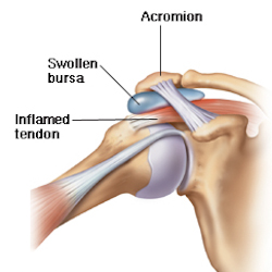
Shoulder Ligament Anatomy Diagram : Equine Reciprocating Systems Examining The Shoulder To Thorax Junction American Farriers Journal : A joint capsule is a watertight sac that surrounds a joint.
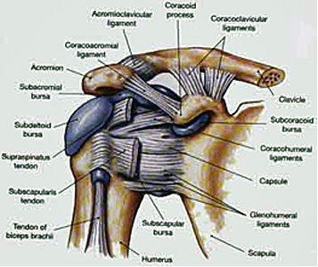
Shoulder Ligament Anatomy Diagram : Equine Reciprocating Systems Examining The Shoulder To Thorax Junction American Farriers Journal : A joint capsule is a watertight sac that surrounds a joint.. There are five major shoulder ligaments that keep the shoulder in place and prevent it from dislocating. All about the shoulder muscles. This acts as the bony framework by which the muscles of the chest, upper back and shoulder connect the upper limb to the trunk of the body and control it's movements.the clavicle connects to the sternum via the. The primary function of the shoulder girdle is to give strength and range of motion to the arm. Notice superior labrum and attachment of the superior glenohumeral ligament.
Related online courses on physioplus. The shoulder joint (glenohumeral joint) is a ball and socket joint between the scapula and the humerus. The conoid and trapezoid ligaments make up the coracoclavicular ligaments. Webmd's shoulder anatomy page provides an image of the parts of the shoulder and describes its function, shoulder problems, and more. The disk has a great variation in size and shape.
Shoulder Tendinitis Causes Treatment Prevention from my.clevelandclinic.org All about the shoulder muscles. Static:gh ligaments, labrum & capsule and dynamic constraints: Ligaments appear as crisscross bands that attach bone to bone and help stabilize joints. The transverse humeral ligament is not shown on this diagram. Home > blog > anatomy > shoulder anatomy: Roots, trunks, divisions, cords, branches, clinical anatomy. The conoid and trapezoid ligaments make up the coracoclavicular ligaments. Divided into two additional ligaments including the trapezoid ligament.
Coronal section of shoulder joint.
It can help you understand our world more detailed and specific. Comprising of numerous ligamentous and muscular structures, the only actual bony articulations are the glenohumeral joint and the acromioclavicular jo. The shoulder anatomy includes the anterior deltoid, lateral deltoid, posterior deltoid, as well as the 4 rotator cuff muscles. Although the joint is held together by these extensive ligament and muscle attachments, certain types of forces can weaken the shoulder easily. Ligaments appear as crisscross bands that attach bone to bone and help stabilize joints. This page is about shoulder anatomy ligaments and muscles,contains soft tissues of the shoulder,shoulder joint; Understanding shoulder anatomy can help to avoid injury, promote rehabilitation, and can assist you in using the joint optimally. The human shoulder is made up of three bones: Roots, trunks, divisions, cords, branches, clinical anatomy. Ac joint is a diathrodial joint with a fibrocartilaginous disk. The disk has a great variation in size and shape. Related online courses on physioplus. Last update february 25, 2021.
It can help you understand our world more detailed and specific. The shoulder is not a single joint, but a complex arrangement of bones, ligaments, muscles, and tendons that is better called the shoulder girdle. These ligaments are the primary restraint for upward and backward movement of the. A joint capsule is a watertight sac that surrounds a joint. Anatomy is the amazing science.
Ligament Physiopedia from www.physio-pedia.com Coronal section of shoulder joint. The transverse humeral ligament is not shown on this diagram. Last update february 25, 2021. Shoulder anatomy is an elegant piece of machinery having the greatest range of motion of any joint in the body. Ligaments appear as crisscross bands that attach bone to bone and help stabilize joints. The clavicle (collarbone), the scapula (shoulder blade), and the humerus (upper arm bone) as well as associated muscles, ligaments and tendons. Robin smithuis and henk jan van der woude. Superior glenohumeral ligament and coracohumeral ligament are the primary restraints to posterior translation with the are flexed, adducted and internally acromioclavicular ligament anatomy.
The shoulder joint (glenohumeral joint) is a ball and socket joint between the scapula and the humerus.
Divided into two additional ligaments including the trapezoid ligament. 7 draw labelled diagram showing the relations of shoulder joint. Understanding shoulder anatomy can help to avoid injury, promote rehabilitation, and can assist you in using the joint optimally. An image depicting shoulder anatomy can be seen below. It is the major joint connecting the upper limb to the trunk. Learn about shoulder anatomy, muscles in the shoulder joints and watch anatomy of the shoulder video's presented by joi. Bones in shoulder, ligaments of the shoulder joint, parts of the shoulder joint, shoulder anatomy, shoulder joints and muscles, shoulder structure anatomy, shoulder tendon anatomy, shoulder related posts of diagram of shoulder muscles and tendons. Diagram of the shoulder anatomy of shoulder ligament ideas anatomy lesson full hd wallpaper. The shoulder is not a single joint, but a complex arrangement of bones, ligaments, muscles, and tendons that is better called the shoulder girdle. Glenohumeral joint,shoulder tendons,8 ejercicios para el hombro que debemos hacer and more. Anatomy is the amazing science. (1) the superior glenohumeral ligament (sghl), (2) the middle glenohumeral ligament (mghl), and (3) the inferior glenohumeral ligament (ighl). Ac joint is a diathrodial joint with a fibrocartilaginous disk.
The shoulder is not a single joint, but a complex arrangement of bones, ligaments, muscles, and tendons that is better called the shoulder girdle. Anatomy is the amazing science. Although three ligaments protect and surround the shoulder joint, most of its stability comes from the powerful muscles and tendons of the rotator cuff. The conoid and trapezoid ligaments make up the coracoclavicular ligaments. Because of its location superior to the glenohumeral joint, it acts as a protection to the joint.
Shoulder Anatomy And Biomechanics Musculoskeletal Key from i2.wp.com Coronal section of shoulder joint. Static:gh ligaments, labrum & capsule and dynamic constraints: You can see it enclosing the glenohumeral joint and you can see its attachment on the anatomical you've got the transverse humeral ligament and the coracohumeral ligament. An image depicting shoulder anatomy can be seen below. It can help you understand our world more detailed and specific. Rotator cuff & scapula stabilising. Bones in shoulder, ligaments of the shoulder joint, parts of the shoulder joint, shoulder anatomy, shoulder joints and muscles, shoulder structure anatomy, shoulder tendon anatomy, shoulder related posts of diagram of shoulder muscles and tendons. The primary function of the shoulder girdle is to give strength and range of motion to the arm.
The disk has a great variation in size and shape.
The transverse humeral ligament is not shown on this diagram. The primary function of the shoulder girdle is to give strength and range of motion to the arm. Ligaments of the shoulder joint (hansen, 2009, pg. (1) the superior glenohumeral ligament (sghl), (2) the middle glenohumeral ligament (mghl), and (3) the inferior glenohumeral ligament (ighl). Anatomy shoulder ligaments tendons pain deltoid shoulder muscle anatomy shoulder bone structure shoulder clavicle anatomy shoulder capsule anatomy shoulder joint model shoulder ligaments diagram shoulder labral anatomy torn shoulder tendon shoulder anatomy. Related online courses on physioplus. Coronal section of shoulder joint. Anatomy is the amazing science. The brachial plexus anatomy animation: The human shoulder is made up of three bones: Static:gh ligaments, labrum & capsule and dynamic constraints: Editor · aug 6, 2017 ·. Diagram of the shoulder anatomy of shoulder ligament ideas anatomy lesson full hd wallpaper.
Robin smithuis and henk jan van der woude shoulder anatomy diagram. This diagram here just shows the joint capsule itself.


0 Komentar